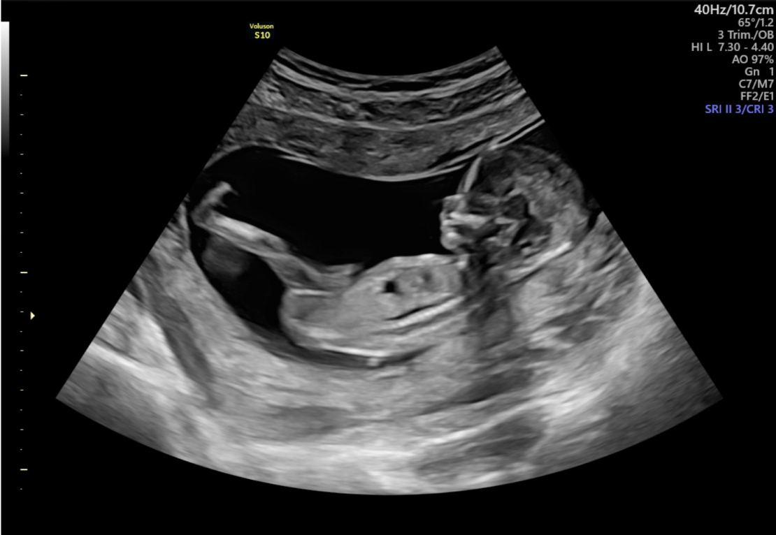
Making ultrasounds, the most beautiful and best images of your little one
Throughout your pregnancy, we will conduct regular ultrasound examinations. While you and your partner enjoy the view of your baby, the sonographer will assess your baby’s growth, development and position. We will also monitor the physical contours and organ structure.
Ultrasounds at Femme Amsterdam
During your pregnancy you will receive at least 8 ultrasounds at Femme Amsterdam. In the Netherlands, you are offered 3 ultrasounds as standard, which are also reimbursed by your insurance or from a subsidy from the Ministry of Health, Welfare and Sport. These are the term ultrasound, the 13 and 20 week ultrasound.
At Femme Amsterdam we believe that through multiple ultrasounds we can better monitor the growth and development of your baby. See the timeline below for all ultrasounds that you can get in practice at Femme Amsterdam.
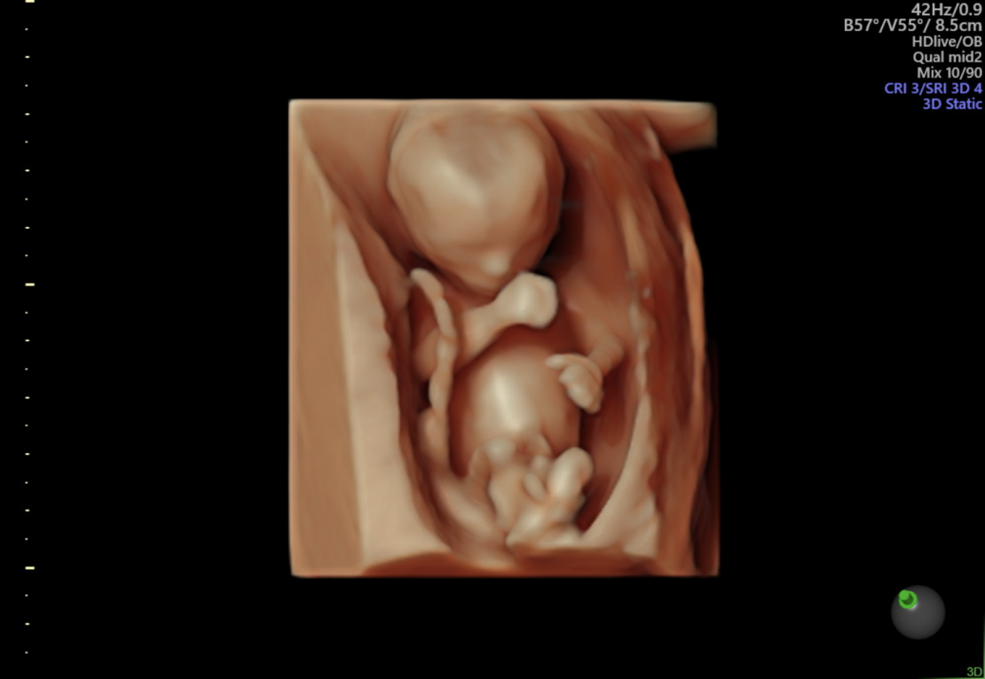
If you like, we will also look at your baby in 3D and 4D every ultrasound.
Afterwards you will receive the photos printed out and we will send photos and videos digitally to your phone.
Check out our echo timeline below.
echo timeline
You receive these ultrasounds at Femme Amsterdam
-
Early ultrasound | around 7th or 8th weeks
An early ultrasound, also called a vitality ultrasound, is made around the 7th or 8th week of your pregnancy. During this ultrasound, our midwife checks whether the heart is beating and whether the fertilized egg has implanted itself in the right place in the uterus (not ectopic). We also check whether there are multiple births. The early ultrasound is an internal, vaginal ultrasound. That is why we advise you to urinate beforehand.
- Duration: 20 min
- Cost: This echo is included in our Basic+, Excellent and Premium package. A separate early ultrasound costs €65. (Sometimes there are clear reasons to perform an early ultrasound. In that case, the ultrasound will be reimbursed by your health insurer.)
-
Due date scan | around 10 weeks
During the term ultrasound, we measure the length of your baby and determine when you are due. We listen to the heart and look at the body contours, such as the hands and feet.
- Duration: 20 minutes
- Costs: This ultrasound is reimbursed by your health insurer
-
First trimester SEO | 13 week ultrasound
The 13-week ultrasound is an ultrasound examination for physical abnormalities. The child is still small, but some, often serious, abnormalities can be seen. If you opt for the 13-week ultrasound, you are taking part in a scientific study; the IMITAS study. In this study, the sex of the child may not be disclosed. The ultrasound may take place between a pregnancy of 13 and 14 weeks and three days. If you are on holiday just during that period, discuss the other options with us.
Duration: 20-30 minutes
Costs: This ultrasound is fully reimbursed from a subsidy from the Ministry of VSW. -
Gender determination | around 13/14 weeks
Are you curious about the gender of your baby? From week 13/14 your child is completely complete. In this fun ultrasound we can look at the size and gender of your child.
- Duur: 20 minuten
- Kosten: This ultrasound is included in our Excellent and Premium package. A separate gender determination ultrasound costs €65.
-
Structural Ultrasound Examination | 20 week ultrasound
The 20 week ultrasound is intended to examine the health of your baby. The ultrasound is also called Structural Ultrasound Examination. During this ultrasound we look extensively at the development of the organs and any structural abnormalities can be detected. Such as an open back, heart defect or an open skull. This ultrasound can be performed between 18-20 weeks.
- Duration: 40 to 50 minutes
- Costs: This ultrasound is reimbursed by your health insurer.
-
Fun ultrasound full-colour | starting at 24 weeks
From about 24 weeks you can have a fun ultrasound made in color. With this ultrasound you can see your baby in perspective and in color. The 3D echo is made up of several 2D images. A 4D echo is a moving 3D echo in color and is recorded ‘live’.
- Duration: 20 to 30 minutes
- Cost: The fun echo in color is included in our Basic+, Excellent and Premium package. A separate fun ultrasound costs €99,95
-
Growth ultrasounds | from 24 weeks
During a growth ultrasound we measure the size of the head; head circuference, the waist circumference and the upper leg length, this combination gives an estimated fetal weight. We place all this data in a graph so that we can accurately follow the growth line of your child. We also check the amount of amniotic fluid.
- 24 weeks | Your baby is now on average 28 cm and weighs on average 650 grams
- 28 weeks | Your baby is now on average 31 cm and weighs on average 1400 grams
- 31 weeks | Your baby is now on average 35 cm and weighs on average 1700 grams. In addition to checking the growth and amniotic fluid, we also check whether your placenta is far enough from the exit.
- 33 weeks | Your baby is now on average 38 cm and weighs on average 2100 grams.
- 35 weeks | Your baby is now on average 41 cm and weighs on average 2500 grams. We now also look closely at the position of your child. 95% of all babies are now head down.
- 37 weeks | Your baby is now on average 44 cm and weighs on average 2900 grams.
- 38 weeks | Your baby is now an average of 47 cm and weighs an average of 3100 grams
- 39 weeks | Your baby is now on average 49 cm long and weighs on average 3300 grams
- 40 weeks | Your baby is now on average 51 cm long and weighs on average 3500 grams
- 41 weeks | Your baby is now on average 54 cm and weighs on average 3800 grams.
- Duration: 20 minutes
- Cost: The growth ultrasounds are included in our Basic+, Excellent and Premium package. A separate growth ultrasound costs €65.
frequently asked questions
Over het maken van echo’s
Yes, that is fine. Do ensure that the oil is well absorbed before the ultrasound begins. Unabsorbed oil prevents the sound waves from effectively penetrating the abdomen resulting in a blurred image.
No, your baby is not affected by ultrasound imaging. Babies cannot hear the ultrasound waves.
An ultrasound cannot detect mental or genetic disorders. The chance of discovering physical congenital abnormalities depends on the specific disorder. Research shows that there is a 90% chance of identifying spina bifida and approximately 25-50% for heart defects depending on severity. The bigger the baby gets the easier disorders are detected,
No, an ultrasound exam cannot cause a miscarriage.
Having multiple ultrasound scans during your pregnancy allows our midwives to monitor the growth of your baby. A potential growth deceleration can be detected. External examinations (feeling the abdomen) only detect slow fetal growth in approximately 50% of cases.
Ultrasounds have been used in the medical world for many years and are not harmful to the mother or baby.
An ultrasound is made using ultrasonic sound waves. These sound waves pass through your abdomen and bounce off the fetus providing body tissue an image on the ultrasound screen.